
This article is for informational purposes only.
“He always coughs and struggles to breathe after exercise, but whenever we test him, everything seems normal."
This is one of the most common reasons children are referred to pulmonologists and allergists. Symptoms occurring during physical activity - such as shortness of breath, cough, or wheezing - may suggest exercise-induced bronchoconstriction. The challenge is that standard diagnostic tests do not always confirm the diagnosis.
In these situations, physicians often suspect exercise-induced bronchoconstriction (EIB), a temporary narrowing of the airways that occurs during or after physical activity.
For many years, the primary test used to confirm this diagnosis has been spirometry. However, clinicians have long observed an intriguing paradox: some children experience obvious wheezing and shortness of breath after exercise despite having normal spirometry results. Conversely, some children with abnormal lung function tests do not produce any characteristic respiratory sounds.
Could wheezing and rhonchi after exercise reflect physiological processes that conventional spirometry cannot capture?
Researchers from Sapienza University in Rome set out to answer this question by combining artificial intelligence, digital auscultation, and advanced lung function testing.
Exercise-induced bronchoconstriction is a temporary narrowing of the airways that occurs during or after physical activity. It may affect both patients with asthma and individuals without a previously diagnosed respiratory disease. The most common symptoms include cough, shortness of breath, wheezing, and chest tightness.
Because these symptoms can have many different causes, symptom reporting alone is not sufficient to establish the diagnosis. For this reason, physicians perform an exercise challenge test. This usually involves intensive exercise on a treadmill followed by repeated assessments of lung function. If lung function deteriorates sufficiently after exercise, exercise-induced bronchoconstriction can be confirmed.
Spirometry is the most widely used pulmonary function test performed in pulmonology and allergy clinics. It requires patients to take a deep breath and then exhale as forcefully and rapidly as possible into a device that measures airflow through the airways.
Spirometry plays a crucial role in the diagnosis of asthma and other obstructive lung diseases. However, it has certain limitations. It requires active patient cooperation, making it difficult to perform in younger children, especially preschoolers. In addition, spirometry primarily reflects the function of larger airways, while more subtle abnormalities involving the small airways may remain undetected.
Oscillometry is a much less familiar pulmonary function test. Unlike spirometry, it does not require forced breathing maneuvers. Instead, patients simply breathe quietly through a mouthpiece while the device analyzes the propagation of small pressure oscillations through the respiratory system.
This allows oscillometry to assess the mechanical properties of the lungs, including the function of the small airways, which are increasingly recognized as an important component of the pathophysiology of asthma and exercise-induced bronchoconstriction.
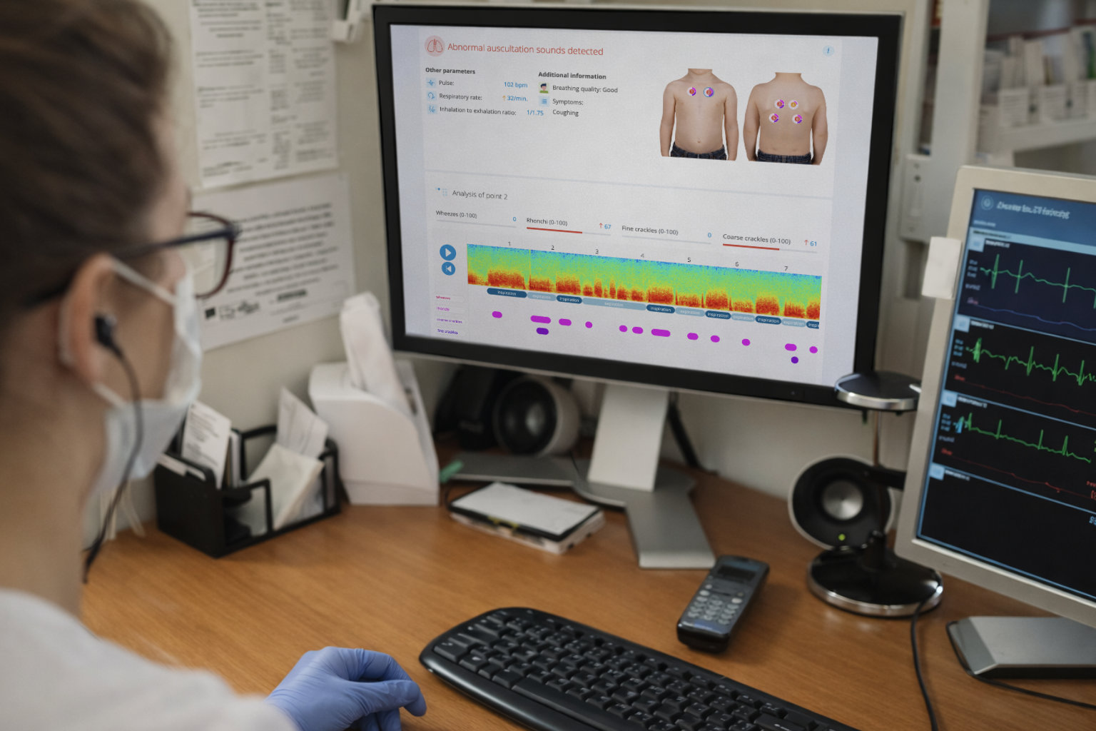
Artificial intelligence-assisted digital stethoscopes represent one of the newest technologies being developed in respiratory medicine. Devices such as StethoMe can automatically detect and analyze abnormal respiratory sounds, including wheezes and rhonchi, which occur during airway obstruction.
The examination is fast, non-invasive, and can be performed both in clinical settings and at home. By enabling objective and repeatable analysis of respiratory sounds, AI-assisted digital auscultation is attracting growing interest as a novel approach to respiratory diagnosis and monitoring.
Researchers from Sapienza University in Rome sought to determine whether abnormal respiratory sounds detected by artificial intelligence after exercise reflect actual physiological changes occurring in children's airways. In particular, they wanted to investigate whether wheezes and rhonchi recorded by an AI-assisted digital stethoscope were associated with the results of conventional pulmonary function tests.
The study included 89 children reporting recurrent respiratory symptoms during physical activity. All participants underwent a standardized treadmill exercise challenge followed by spirometry, oscillometry, and respiratory sound analysis using the StethoMe digital stethoscope supported by artificial intelligence algorithms. Exercise-induced bronchoconstriction was confirmed in 21 children based on spirometric criteria. The researchers then compared the presence of wheezes and rhonchi detected by AI with the results of pulmonary function testing.
The first important observation was that artificial intelligence showed excellent agreement with physician assessments. Agreement between the digital stethoscope and clinicians was very high for wheezes (κ=0.92) and high for rhonchi (κ=0.77). For comparison, a kappa value of 1 indicates perfect agreement, meaning that AI and physicians identified the same abnormal respiratory sounds in the vast majority of cases. Moreover, in several cases, the AI system detected pathological respiratory sounds that were not recognized during conventional auscultation.
However, the most interesting findings emerged when respiratory sounds were compared with pulmonary function tests.
Children who developed wheezes or rhonchi after exercise did not differ significantly in spirometry results from children without abnormal respiratory sounds. In other words, the presence of wheezing after exercise did not reflect the results of conventional lung function testing.
At the same time, these children showed clear abnormalities on oscillometry. The strongest associations involved parameters describing expiratory small airway function. Furthermore, the greater the intensity of wheezes and rhonchi detected by artificial intelligence, the more abnormal the oscillometry findings were.
The findings of this study suggest that wheezes and rhonchi occurring after exercise may reflect physiological processes taking place primarily in the small airways - an area that conventional spirometry does not fully assess.
This does not mean that artificial intelligence replaces spirometry. Rather, the study suggests that respiratory sound analysis may provide valuable and precise information about respiratory function, particularly when standard pulmonary function tests do not fully explain a patient's symptoms.
The study conducted by Italian researchers demonstrates that combining artificial intelligence, digital auscultation, and advanced pulmonary function testing may help us better understand what actually happens in the airways during exercise. Importantly, respiratory sound analysis may not only support the detection of pathological phenomena but also provide insight into the physiological mechanisms underlying them.
One of the greatest advantages of AI-assisted digital stethoscopes is their ability to monitor patients outside the physician's office — precisely when symptoms occur. Unlike tests performed during a scheduled clinic visit, respiratory sound analysis may provide information about what is happening in the airways under real-life conditions.
Want to monitor your child's breathing between doctor visits? StethoMe detects abnormal lung sounds at home — the same way a doctor does, but available every day.
Learn about StethoMe